Fluorescence Microscope Magnification

Fluorescent Microscopy Images 40 Magnification Of Lncap A And

Gxm Ztxv7 10x 40x Stereo Microscope V7 Stand Gt Vision Online

Fluorescent Microscopy

Fluorescence And Electron Microscopy

Fluorescent Microscopy

Fluorescent Microscopy

But what can you see with a coin microscope.
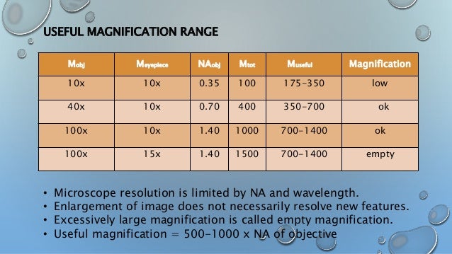
Fluorescence microscope magnification. The range of useful magnification for an objectiveeyepiece combination is defined by the numerical aperture of the microscope optical system. Since x rays penetrate most objects there is no need to specially prepare them for x ray microscopy observations. Instead of white light ie. Welcome to the olympus microscopy resource center designed to provide an internet based educational forum on all aspects of optical microscopy photomicrography and digital imaging.
A key difference in a fluorescence microscope is the light supply. There is a minimum magnification necessary for the detail present in an image to be resolved and this value is usually rather arbitrarily set as 500 times the numerical aperture 500 x na and defined. An x ray microscope uses electromagnetic radiation in the soft x ray band to produce magnified images of objects. When using a high power microscope also known as a compound microscope it is best to start out with the lowest magnification get your specimen in focus and then move up to the higher magnifications one at a time.
The optical microscope often referred to as the light microscope is a type of microscope that commonly uses visible light and a system of lenses to magnify images of small objects. A narrow range of wavelengths. The molecular expressions website features hundreds of photomicrographs photographs through the microscope of everything from superconductors gemstones and high tech materials to ice cream and beer. What will you be able to see under a high power microscope.
Many wavelengths that is used in brightfield microscopes fluorescence microscopes require light of a specific color ie.

Ex Vivo Vessel Tissues Were Imaged With A Fluorescence Microscope At

Microscope

Understanding Microscopes And Objectives Edmund Optics

Fluorescence Microscopist Co Uk

Fluorescence Microscopy
Fluorescence Microscopy Errors Olympus Life Science

Fluorescent Microscopy 400x Magnification Of Vero Cells Infected

Super Resolution Fluorescence Microscopy Ppt Video Online Download
Microscopy

Chapter 2 Observing The Microbial Cell Ppt Download

Hek 293a Cells Infected With Radv By Fluorescence Microscopy And
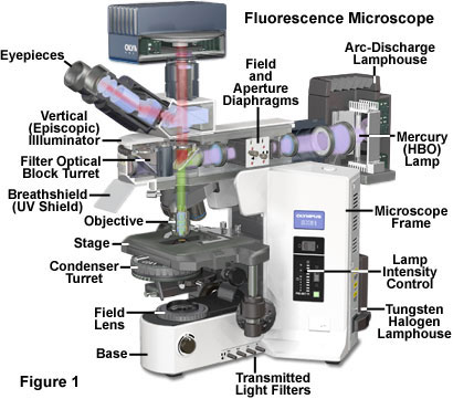
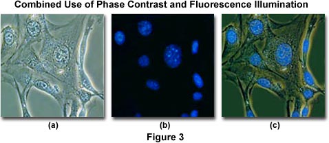
Fluorescence Microscopy Anatomy Of The Fluorescence Microscope

Microscopes Microbiology
