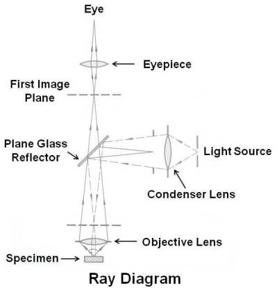
Fluorescence Microscope Ray Diagram

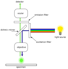
Working Principle Of Fluorescence Microscope With Figure

Fluorescent Microscopy
Fluorescence Microscope Wikipedia

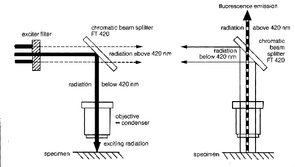
Light Path Of A Confocal Fluorescence Microscope Out Offocus Light

Confocal Microscopy Microscopist Co Uk

Light Path Of A Confocal Fluorescence Microscope Out Offocus Light
Confocal microscopy x ray fluorescence imaging is a newer technique that allows control over depth in addition to horizontal and vertical aiming for example when analysing buried layers in a painting.
Fluorescence microscope ray diagram. However some of them after absorbing light of a particular wavelength and energy emit light of a longer wavelength and lesser energy. Fluorescence is one of the most commonly used physical phenomena in biological and analytical microscopy mainly because of its high sensitivity and high specificity. Many substances absorb light. Jablonski diagram for fluorescence an excited state electron rapidly on the order of 10 12 seconds loses its energy to vibration heat a process called internal conversion and falls to the lowest level of the first s 1 excited state.
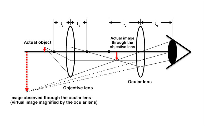
Fluorescence microscopy has become an essential tool in biology as well as in materials science due to attributes that are not readily available in other optical microscopy techniques. A fluorescence microscope is an optical microscope that uses fluorescence and phosphorescence instead of or in addition to scattering reflection and attenuation or absorption to study the properties of organic or inorganic substances. A popular method of representing a train of propagating light waves involves the application of geometrical optics to determine the size and location of images formed by a lens or multi lens system. An introduction to fluorescence spectroscopy 5 luminescence and the nature of light a hot body that emits radiation solely because of its high temperature is said to.
Geometrical construction of ray diagrams a popular method of representing a train of propagating light waves involves the application of geometrical optics to determine the size and location of images formed by a lens or multi lens system. Find out how fluorescence microscopes support your research.

Basic Fluorescence Microscopy Lenstrotek

Top 8 Types Of Microscopy With Diagram

Microscopy

Principle Of Confocal Fluorescence Microscopy Download Scientific

Epifluorescence Microscope Diagram Wiring Diagram
Metallurgical Microscopes Microscopegenius Com

Epifluorescence Microscope Diagram Wiring Diagram

Understanding Microscopes And Objectives Edmund Optics
Epifluorescence Microscope Diagram Wiring Diagram
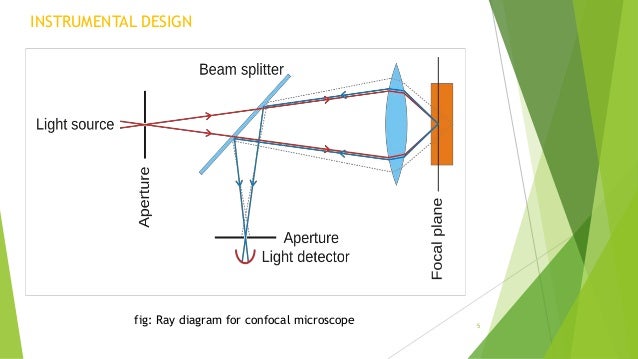
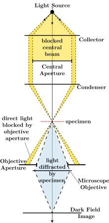
Confocal Microscopy

第二章
Portfolio Avr Optics Inc Part 19
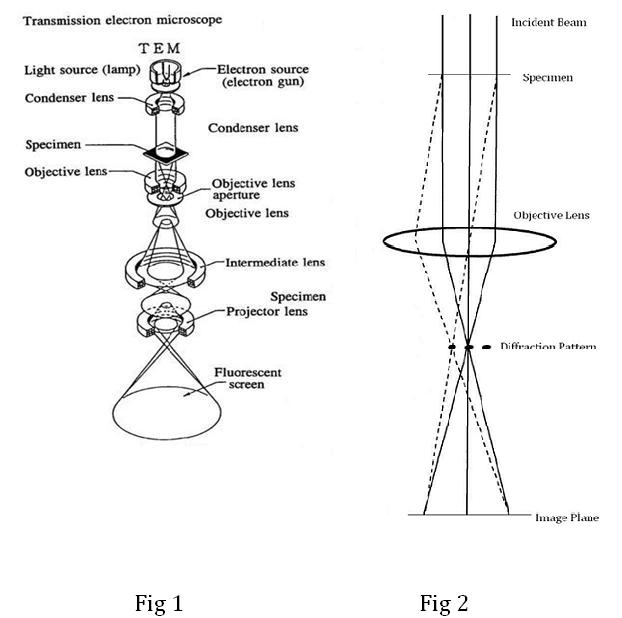
Transmission Electron Microscopy Tem