Fluorescence Microscopy Bacteria

Images Of Bacterial Cells Observed By Using A Fluorescence

As Imaged By Fluorescence Microscopy A The Attachment Of P

Fluorescence Microscopy Images Of Ao Eb Dual Staining Bacterial

Energy Dependent Motion Of Tonb In The Gram Negative Bacterial Inner

Bacteria From Freshwater Ecosystems Seen Under Fluorescence
Viaquant Viability Cytotoxicity Kit For Bacteria Cells Genecopoeia
The light microscope so called because it employs visible light to detect small objects is probably the most well known and well used research tool in biology.
Fluorescence microscopy bacteria. There are three core laboratories on campus that offer expertise instruction and instrumentation in optical microscopy for research. In contrast to phosphorescence the term fluorescence is applied to substances without after glow ie fluorescence ceases directly with the stimulant irradiation. However current microscopy techniques limit the quantity and quality of information available to researchers and clinicians. Optical or light microscopy involves passing visible light transmitted through or reflected from the sample through a single lens or multiple lenses to allow a magnified view of the sample.
Fluorescence is the characteristic phenomenon of luminosity of solids liquids or gases after exposure to light. Light emitting stains such as fluorescently labelled antibodies are applied to samples to enable vizualisation of specific structures. Autofluorescence can be problematic in fluorescence microscopy. Efforts to identify the molecular basis for the novel fluorescence in jellyfish began with osamu shimomuras studies of the aequorea jellyfish in the early 1960s.
Research and diagnosis in the life sciences depends on the information that can be found using cellular analysis. Subsequently analysed using confocal microscopy. Confocal laser scanning microscopy clsm is another powerful tool to visualize polymer gels labeled with fluorescent ligands. The resulting image can be detected directly by the eye imaged on a photographic plate or captured digitally.
For apa capsules alginate can be labeled with the fluorochrome fluoresceinamine and poly l lysine can be labeled with alexa 546 protein labeling kit strand et al 2003. The cnr biological imaging facility this lab.

Bacterial Roundabouts Determine Cell Shape Scientists Decipher

Efficient Gene Transfer In Bacterial Cell Chains Mbio

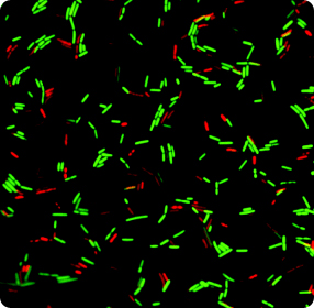
How Can You Tell If Bacteria Are Alive Or Dead Sustainable Nano

Bacteria
Molecular Expressions Microscopy Primer Specialized Microscopy

Fluorescent Antibody Techniques Microbiology

Detection Of Low Copy Number Genomic Dna Sequences In Individual
Molecular Expressions Microscopy Primer Specialized Microscopy

Microbiological Aspects Of Table Olives Intechopen

29 Best Fluorescent Microscopy Images Fluorescence Microscopy

Tracking How Bacteria Compete For Space Human Frontier Science Program

Fluorescence Microscopy Of Menb Strain H44 76 Colonies A Bacteria

Sem Course Images University Of Florida Institute Of Food And
