Fluorescence Microscopy
Fluorescence Microscope Wikipedia

Fluorescent Microscopy

High Throughput Fluorescence Microscopy New Trend

Fluorescence Microscopy The Magic Of Fluorophores And Filters

Fluorescence Microscopy Illustrations

Fluorescence Microscopy Research Groups Imperial College London
The company has specialised in fluorescence microscopy since it introduced the first commercially available led illumination system in 2004.
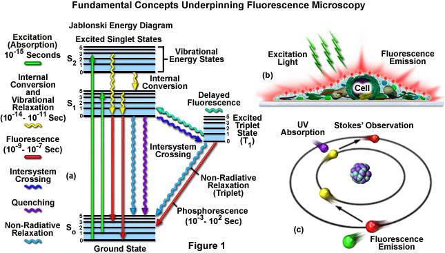
Fluorescence microscopy. First observed in 1560 fluorescence has evolved into a powerful technique that enables entire fields of cutting edge science and medicine. The only spectroscopic technique capable of resolving single molecules fluorescence has moved from the lab to applications limited only by imagination. The olympus microscopy resource center microscopy primer explores many of the aspects of visible light starting with an introduction to electromagnetic radiation. This is because power is usually.
Dedicated to large sample imaging the muvi spim is optimized for long term 3d fluorescence imaging of living specimens such as drosophila larvae and early zebrafish embryos. It can often be difficult to compare the power across. Brukers suite of fluorescence microscopy systems provides a full range of solutions for life science researchers. Coolled designs and manufactures cutting edge led illumination systems for researchers and clinicians using the latest led technology.
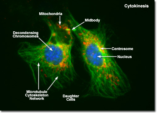
Light sheet fluorescence microscopy lsfm is a fluorescence microscopy technique with an intermediate to high optical resolution but good optical sectioning capabilities and high speed. Light is a phenomenon that is explained with a model based on rays and wavefronts. Observing mitosis with fluorescence microscopy mitosis a phenomenon observed in all higher eukaryotes is the mechanism that allows the nuclei of cells to split and provide each daughter cell with a complete set of chromosomes during cellular division. Horizontal 3600 illumination and detection.

Fluorescence Microscope Wikipedia
Zeiss Microscopy Online Campus Microscopy Basics Fluorescence

Fluorescent Microscopy Lnf Wiki

Fluorescence Microscopy Research Baylor College Of Medicine
Widefield Fluorescence Microscopy What You Need To Know

Fluorescence Microscope Wikipedia

Fluorescence Microscopy Fluorescence Microscope Principle Youtube

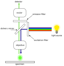
1 The Scheme Of The Fluorescence Microscope Download Scientific
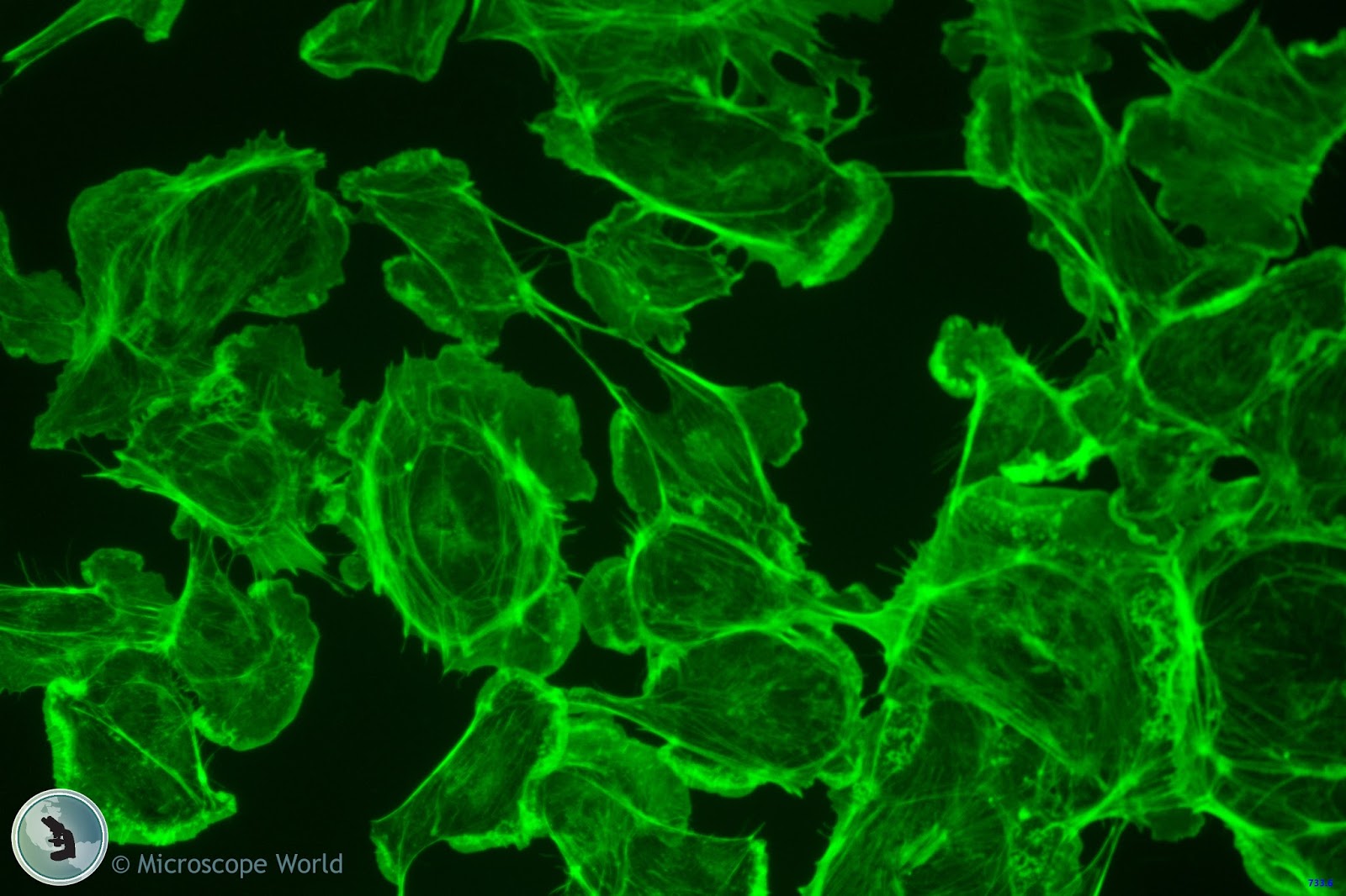
Microscope World Blog Fluorescence Microscopy

Fluorescence Microscopy Illumination Highly Stable Multi Line

Encyclopedia Of Laser Physics And Technology Fluorescence

Charming Fiber Lasers And Components For Fluorescence Microscopy

Epifluorescence Microscope Basics Thermo Fisher Scientific Us
