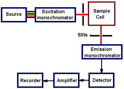
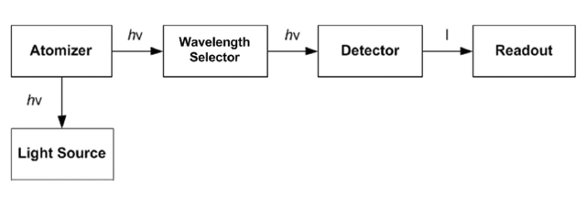
Fluorescence Spectroscopy Block Diagram

4 Schematic Diagram Of A Fluorescence Spectrometer Download

4 Schematic Diagram Of A Fluorescence Spectrometer Download

Absorption And Fluorescence Spectroscopy Of 1 2 3 4
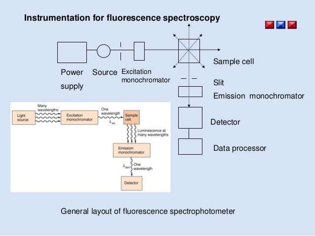
Molecular Fluorescence Spectroscopy

Block Diagram Of A Typical Fluorescence Spectroscopy Download
Uv Vis Luminescence Spectroscopy Instrumentation
Raman spectroscopy is commonly used in chemistry to provide a structural fingerprint by which molecules can be identified.
Fluorescence spectroscopy block diagram. Cancer biomarkers with high selectivity specificity and reproducibility play essential role in diagnosis prognosis and prediction of treatment efficacy in cancer patients. Preparation of standard solution of k 2cr 2o 7. The program includes simple spectroscopy applications such as absorbance transmittance reflectance and radiometry. Raman is a spectroscopic technique used to observe vibrational rotational and other low frequency modes in a system.
Named after indian physicist sir c. Spectroscopy consists of many different applications such as atomic absorption spectroscopy atomic emission spectroscopy ultraviolet visible spectroscopy x ray fluorescence spectroscopy infrared spectroscopy raman spectroscopy nuclear magnetic resonance. Polyethylene oxide polypropylene oxide polyethylene oxide block copolymer surfactants in aqueous solutions and at interfaces. Vba source code program for ms excel to instantly grab graph spectral data from spectrometer.
Each column within each half of the sensor is equipped with dual column level amplifiers and dual analog to digital converters adc represented as a block diagram in below. X ray fluorescence xrf spectrometry is an elemental analysis technique with broad application in science and industry. First year industrial chemistry practical. Xrf is based on the principle that individual atoms when excited by an external energy source emit x ray photons of a characteristic energy or wavelength.
View and download horiba fluorolog 3 operation manual online. Raman spectroscopy r m en. To find out the concentration of unknown k 2cr 2o. Raman hr tec x2 405 532 633 785 830 high resolution configuration with high sensitivity detector lens assembly 16 bit low noise electronics high resolution slit and 2 stage thermoelectric detector cooler.
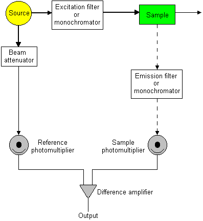
Bio Medical Engineering Solutions Sl Atomic Fluorescence Spectroscopy

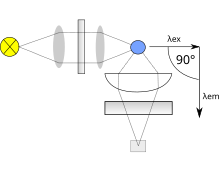
Block Diagram Of Atomic Fluorescence Spectrometry Chemistry

Block Diagram Of A Typical Fluorescence Spectroscopy Download
Fluorescence Spectroscopy Wikipedia
Determination Of Mercury In The Environment Petrochemical Products
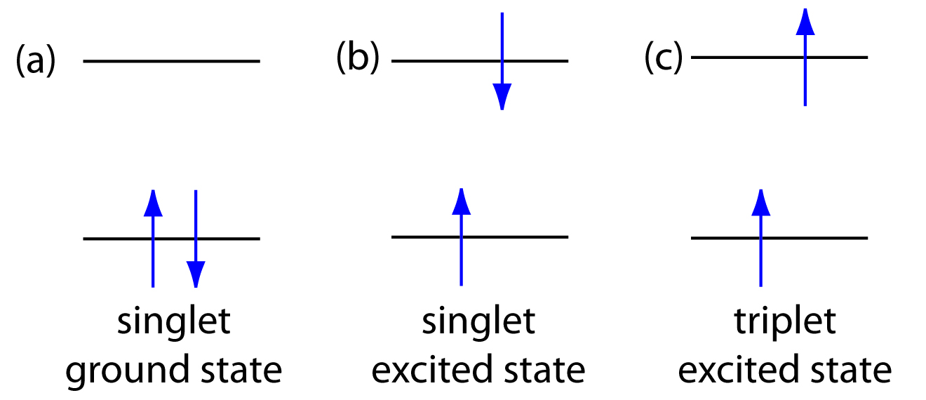
Introduction To Fluorescence And Phosphorescence
Fluorimetry

Fluorescence Spectroscopy Ppt Video Online Download

10 6 Photoluminescence Spectroscopy Chemistry Libretexts

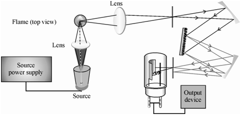
Atomic Absorption And Emission Analysis

Detection Of Ageing High Voltage Electrical Cables Using
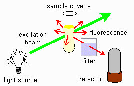
Fluorescence Spectrometry Fluorescence Spectrometry Chromedia
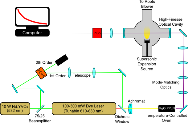
Mccall Research Group Supersonic Source
