Fluorescent Microscope Pictures
Fluorescence Microscope Wikipedia

Fluorescent Microscopy

Fluorescent Microscope At Rs 145000 Piece Fluorescent Microscope

Meiji Techno Mt6000 Epi Fluorescence Led Microscope

Fluorescence Microscope Wikipedia

Fluorescence Microscopy Fluorescence Microscope Principle Youtube

Neurons blood vessels mitochondria etc are visualized because fluorescent compounds attach to only those specific structures.
Fluorescent microscope pictures. Leica mz6 stereo microscope 063x 4x 10x eye lenses 10x objective lens focus mount used but in great working condition some small fractures on case but no scratches on lenses. The fluorescent rocks displayed below were collected from a single mine in the mountains of southern arizona. Fluorescent minerals from southern arizona. Lb 200 binocular biological microscope with finite semi plan achromatic objectives and extra wide field anti fungus with.
Advanced digital photomicrography abstract. Bypassing abbes physical limit of 02 micrometres means that the optical microscope can now peer into the nanoscopic world. Home resources advanced digital photomicrography. In most cases the emitted light has a longer wavelength and therefore lower energy than the absorbed radiation.
It is a form of luminescence. Fluorescence microscopy is an imaging modality used to visualize specific structures in biological and other physical samples. The availability of high quality digital consumer cameras at relatively low prices has made photography with the microscope significantly easier than with traditional film. The objects of interest in the sample eg.
Quantitative fluorescent in situ hybridization q fish is a cytogenetic technique based on the traditional fish methodology. Accu scope 3055 series stereo microscopes. Commenting on the announcement prof thomas barton president of the. In q fish the technique uses labelled cy3 or fitc synthetic dna mimics called peptide nucleic acid pna oligonucleotides to quantify target sequences in chromosomal dna using fluorescent microscopy and analysis software.
Zeiss Microscopy Online Campus Microscopy Basics Fluorescence

Optika Fluorescence Microscope Rs 350000 Piece M S Swift
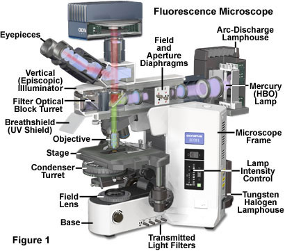
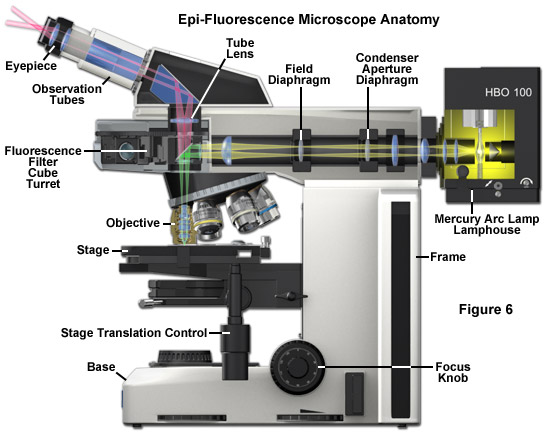
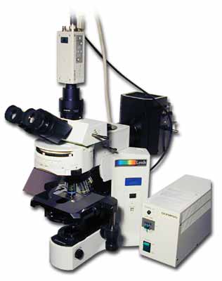
Fluorescence Microscopy Anatomy Of The Fluorescence Microscope
Nikon Eclipse E600 Fluorescence Microscope Uv Dapi Fitc Tritc

Fluorescence Microscopes Biocompare Com

Fluorescence Microscopy Principle And Applications Ppt Download

Fluorescent Microscope Sahmri

Setting Up And Adjusting A Fluorescent Microscope For Reliable

Weswox Research Fluorescent Microscope Fm 5000 Rs 670000 Piece
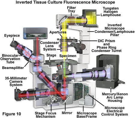
Fluorescence Microscopy Anatomy Of The Fluorescence Microscope
Virtual Phase Contrast And Fluorescent Microscope Wartburg College

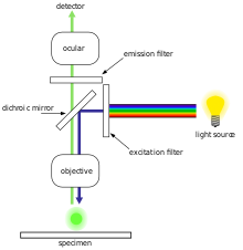
Working Principle Of Fluorescence Microscope With Figure
Motic Fluorescent Microscope Metallurgical Lab Microscopes Bio
