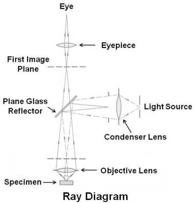
Fluorescent Microscope Ray Diagram

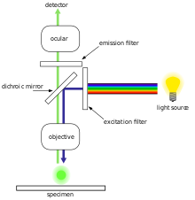
Working Principle Of Fluorescence Microscope With Figure

Fluorescent Microscopy
Fluorescence Microscope Wikipedia

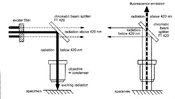
Light Path Of A Confocal Fluorescence Microscope Out Offocus Light

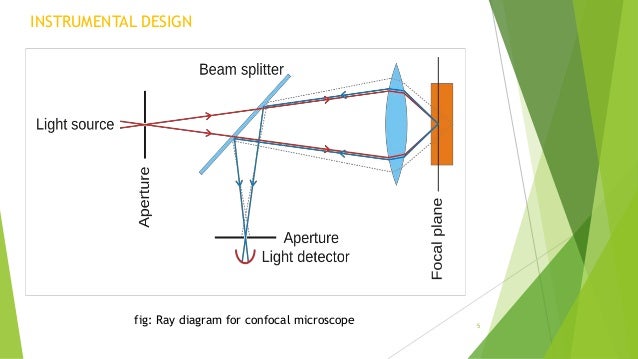
Confocal Microscopy Microscopist Co Uk

Principle Of Confocal Fluorescence Microscopy Download Scientific

Fluorescence microscopy is a special form of light microscopy.
Fluorescent microscope ray diagram. These diagrams clearly explain the functioning of the microscopes along with their respective parts. In practice microbes are stained with a fluorescent dye and then illuminated with blue light. To better understand the structure and function of a microscope we need to take a look at the labeled microscope diagrams of the compound and electron microscope. It uses fluorescence to highlight structures in fixed and living biological specimens instead of using absorption phase or interference effects.
Such substances are called fluorescent substances. Colocalization and interaction studies can be performed and ion concentrations as well as intra and intercellular processes like endocytosis and exocytosis can be observed. X ray fluorescence xrf is the emission of characteristic secondary or fluorescent x rays from a material that has been excited by bombarding with high energy x rays or gamma rays. To meet these requirements we offer high precision fluorescent microsphere reference standards for fluorescence microscopy and flow cytometry and a set of ready made fluorescent standard solutions for spectrofluorometry fluorescence microscopy accessories and reference standardssection 231 flow cytometry reference standardssection 232.
With the help of. Introduction to fluorescence microscopy the absorption and subsequent re radiation of light by organic and inorganic specimens is typically the result of well established physical phenomena described as being either fluorescence or phosphorescence. Fluorescence microscopy even allows users to determine the distribution of a single molecule species its amount and its localization inside a cell. Introduction to fluorescence and the jablonski diagram.

Top 8 Types Of Microscopy With Diagram

Microscopy

Light Path Of A Confocal Fluorescence Microscope Out Offocus Light

Basic Fluorescence Microscopy Lenstrotek

Epifluorescence Microscope Diagram Wiring Diagram
Metallurgical Microscopes Microscopegenius Com

Epifluorescence Microscope Diagram Wiring Diagram
Epifluorescence Microscope Diagram Wiring Diagram
Portfolio Avr Optics Inc Part 19

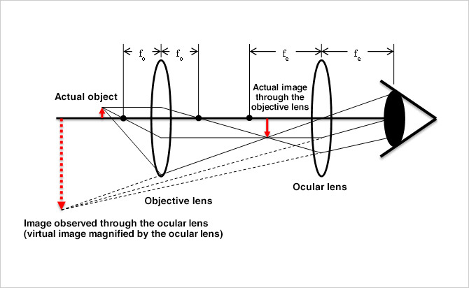
Understanding Microscopes And Objectives Edmund Optics
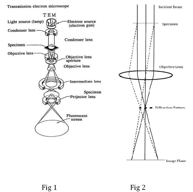
Transmission Electron Microscopy Tem

第二章
Basic Structure And Principle Of Microscopes Keyence Biological