Fluorescent Microscope Resolution

Resolution In Conventional Fluorescence Microscopy Notes A Light

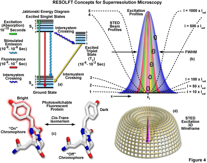
A New Molecular Tool For Continuous Super Resolution Fluorescence
Zeiss Microscopy Online Campus Introduction To Superresolution

A Guide To Super Resolution Fluorescence Microscopy Jcb

Challenges And Trade Offs In Super Resolution Fluorescence
Confocal Microscopy Resolution And Contrast In Confocal Microscopy

Preparing a sample for the scanning electron microscope sem sem preparation.
Fluorescent microscope resolution. Placement on an sem stub sputter coating and insertion into the instrument. Biological samples are often treated with fluorescent dyes to make selected objects visible. Confocal microscopy provides the capacity for direct noninvasive serial optical sectioning of intact thick living specimens with a minimum of sample preparation as well as a marginal improvement in lateral resolution. Microscope cell staining is a technique used to enable better visualization of cells and cell parts under the microscope.
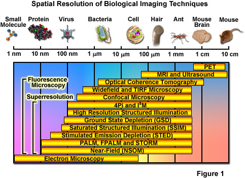
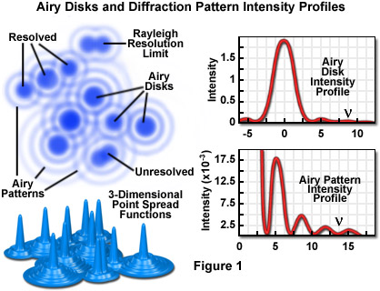
The molecular expressions website features hundreds of photomicrographs photographs through the microscope of everything from superconductors gemstones and high tech materials to ice cream and beer. Superresolution is an emerging technique that holds significant promise for imaging. A fluorescence microscope is an optical microscope that uses fluorescence and phosphorescence instead of or in addition to scattering reflection and attenuation or absorption to study the properties of organic or inorganic substances. The light microscope has a limit of resolution of about 200 nm 02 um.
The quality of the microscopes exceeds that of most other vendors when comparing at similar price levels. Cells observed under a light microscope can be alive or fixed and stained. Download the latest review articles on fluorescent protein technology. 1 fluoviewfrom olympus is open fluoviewmore advanced than ever the olympus fluoview fv1000 confocal laser scanning microscope delivers efficient and reliable performance together with the high resolution required for.
This video walks through the procedures used to prepare samples for electron imaging including.

Fluorescence Microscope Wikipedia
Fluorescence Microscope Wikipedia

Macromolecular Scale Resolution In Biological Fluorescence

Fluorescence Microscopy Nobel Prize Honors Super Resolution

4 Microscopes To Improve The Resolution Confocal And Fluorescent
Zeiss Microscopy Online Campus Introduction To Superresolution

Fluorescence Cryo Microscopy Current Challenges And Prospects

Principles Of Super Resolution Fluorescence Structural Imaging
Bringing Superresolution To Fluorescence Microscopy Features May
Active Motif Super Resolution Microscopy Introduction
Zeiss Microscopy Online Campus Introduction To Superresolution

Researchers Achieve Ultimate Resolution Limit In Fluorescence Microscopy

Multicolor Super Resolution Fluorescence Imaging Imaging
